Fracture Risk in Human Bones
Femoral and humerus fractures are common in the elderly population due to osteoporosis, and are often initiated by falling on the side or on an out-stretched arm. Quantifed computed tomography (QCT), combined with empirical data obtained by biomechanical experiments and computational mechanics may provide the means to enhance our understanding of this frequent injury. The central goal of this interdisciplinary project is the prediction of fracture risk by considering the interplay between micro-mechanical and macroscopic bone models, validated by experiments.
| Project team leader | Dr.-Ing. Stefan Kollmannsberger (TUM), |
| Doctoral Researchers |
Lisa Hug (TUM), Nina Korshunova (TUM) |
| Principal Investigators | Prof. Dr. rer.nat. Ernst Rank (TUM), Professor Zohar Yosibash (Tel-Aviv University) |
| Funding | International Graduate School of Science and Engineering (IGSSE) |
State of the Art
Using CT-scans of human bones it is possible to perform patient-specific finite element simulations in order to predict the strains within the bones. In [1], experiments on fresh frozen proximal humeri were compared to a p-finite element analyses performed on basis of CT scans, where predicted strains in the elastic range corresponded very well with experimental results. Using a simplified maximal principle strain approach, the fracture load could be predicted with 10-15% accuracy. However, up to this day determination of reliable fracture criteria as well as the numerical prediction of fracture initiation and propagation remains a challenge.
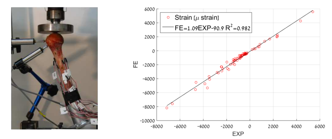
Figure 1: Experimental setup with typical loading and positioning of strain-gauges (left) and correlation to FE-simulation (right) [1].
Phase-Field Modeling
In the field of crack-propagation phase field models have recently shown very promising results. A phase field model smoothes the crack and approximates the discontinuity by introducing a scalar variable, the phase-field, which interpolates between the unbroken and broken states of the material. As the evolution of the crack directly follows from the solution of a partial differential equation, there is no need in complicated and expensive tracking of crack propagation. At the chair, a phase-field model for fracture based on the fnite cell method (FCM) has already been developed and successfully applied in two dimensions [2]. Combining the phase-field approach with FCM represents a promising approach for the simulation of fracture initiation and propagation in human bones.
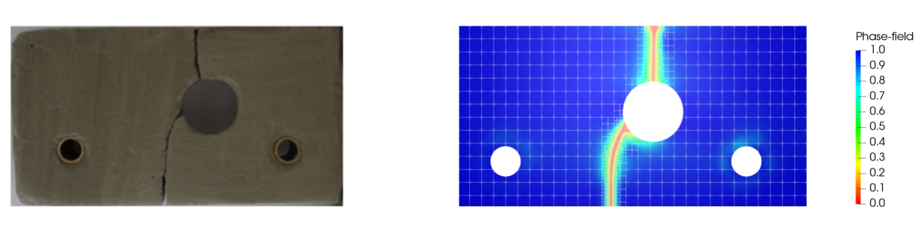
Figure 2: Notched plate with hole. Crack pattern obtained from the experiment (left) corresponds well with FCM simulation result (right) [2].
Objective
In this project, novel numerical fracture initiation and propagation models based on a phase field approach and the finite cell method will be developed. Central goal is the patient-specific prediction of fracture risk which takes into account micro-mechanical properties of the bone.
References
[1] G. Dahan, N. Trabelsi, O. Safran, Z. Yosibash, Verified and validated finite element analyses of humeri, J. Biomech. 49 (2016) 1094–1102. doi:10.1016/j.jbiomech.2016.02.036.
[2] S. Nagaraja, et al. "Phase-field modeling of brittle fracture with multi-level hp-FEM and the finite cell method." arXiv preprint arXiv:1804.08380 (2018).